Research Article - (2023) Volume 12, Issue 6
Introduction: Clinic however, the possible mechanism of bone protection mechanism of Zuogui pill on osteoporosis is still largely unknown.
Methods: An osteoporosis model of postmenopausal breast cancer was generated by gavage of letrozole in mice with ovariectomized breast cancer. Female SPF BALB/c mice were divided into the normal group, pseudocastrated group, model group (letrozole), alendronate group (alendronate tablets) and lowdose, medium-dose and high-dose Zuogui pill groups. Each group was given gavage for 4 weeks according to the corresponding administration plan. Blood was collected from the eyeballs. Serum oestradiol (E2), Bone Alkaline Phosphatase (BALP) and amino terminal propeptide of type Ⅰ collagen (PINP) were detected by Enzyme-Related Immunosorbent Assays (ELISAs). After death, the right femur and tibia were taken and stained with he to observe the bone histopathology. Microcomputed Tomography (μCT) was used to detect bone density and trabecular microstructure in vitro. The protein expression levels of Wnt3a, β-catenin and Runx2 in bone tissue were detected by Western blots.
Results: Compared with the model group treatment, Zuogui pill significantly decreased the serum level of Bone Alkaline Phosphatase (BALP) (P<0.01) and the level of amino terminal Procollagen Ⅰ Propeptide (PINP) (P<0.01) but had no significant effect on oestradiol (E2) (P>0.05). Zuogui pill improved bone tissue morphology, bone microstructure and bone mineral density and the degree of improvement was similar to that of the alendronate group. Compared with those of the model group, the protein expression levels of Wnt3a, β-catenin and Runx2 in the high-dose, middle-dose and low-dose Zuogui pill groups and alendronate sodium groups were significantly increased (P < 0.05).
Conclusion: Zuogui pill has a bone protective effect through the Wnt/β-catenin and Wnt/Runx2 pathways and has good application value in the treatment of osteoporosis.
Zuogui pill • Endocrine therapy • Osteoporosis • Wnt/β- catenin signalling pathway • Wnt/Runx2 signalling pathway
E2: Serum Oestradiol; BALP: Bone Alkaline Phosphatase; PINP: Amino Terminal Propeptide of Type I Collagen; PMOP: Postmenopausal Osteoporosis; AIs: Aromatase Inhibitors; BMD: Bone Density; BV/ TV: Bone Volume fraction; Tb.Th: BONE Trabecular Thickness; Tb.N: Bone Trabecular Number; Tb.Sp: Bone Trabecular Separation; OC: Osteoclasts; OB: Osteoblasts; OP: Osteoporosis; TCF: T-Cell-Specific Transcription Factor; LEF-U: Lymphoid Enhancing Factor
The incidence of breast cancer ranks first among female malignant tumours, accounting for nearly a quarter of all female cancers and the incidence rate (11.7.3%) is the highest among all cancers and death rate is 13.0%, which seriously endangers women's physical and mental health. Breast cancer occurs more frequently in postmenopausal women and its incidence is on the rise globally. Ovarian function declines after menopause and oestrogen levels fall in the hypothalamus-pituitary-ovarian axis, affecting bone metabolism and producing Postmenopausal Osteoporosis (PMOP). In postmenopausal breast cancer patients, oestrogen mainly comes from tissues outside the ovary and is formed by aromatization of androstenedione and testosterone. Aromatase is an essential substance in this link and Aromatase Inhibitors (AIs) can inhibit the activity of this enzyme and block the synthesis of oestrogen [1]. To control the growth of breast cancer cells and treat breast cancer, researchers widely use AIs in the treatment of postmenopausal breast cancer patients. Endocrine therapy for breast cancer and physiological characteristics after menopause are both important risk factors for osteoporosis. Relevant studies have shown that endocrine therapy with AIs can effectively improve the survival rate of breast cancer patients but will increase the occurrence of osteoporosis and pathological fracture. Wu Peili et al. showed that the prevalence of osteoporosis in patients with breast cancer after endocrine therapy reached 40.8%, which reduced the quality of life of patients [2].
Clinical postmenopausal breast cancer patients after endocrine (AL) treatment are prone to secondary osteoporosis, manifested by extreme bone density reduction, waist and leg pain, limb weakness, humpback, easy fracture and other symptoms, with high incidence, high disability and high mortality rates and other characteristics, seriously affecting the quality of life of patients. This condition results in a heavy economic burden to the family and society. Currently, the antiosteoporosis drugs commonly used in the clinic are mainly bisphosphonates and calcitonin, which have many issues, such as many side effects, high cost, risk of drug dependence, risk of kidney damage and low compliance. Therefore, safe, effective, low-cost and tolerable treatment methods are still lacking clinically [3].
Traditional Chinese medicine has good advantages in treating this disease. Chinese medicine states that "bone bi", "bone impotence" and kidney deficiency are the core pathogeneses of this disease, mainly based on the basic theory of traditional Chinese medicine called "kidney main bone" and the rise and fall of the kidney qi determines whether the bone growth and development is strong [4]. In postmenopausal breast cancer patients, the kidney essence is weak and the marrow source is not sufficient to nourish the bone. Liver and kidney are mutual resources and kidney deficiency also results in liver deficiencies, which prevents Rongjin bundle benefits for bones and joints; thus, Zuogui pill is used for the liver and kidney to strengthen bones and muscles for treatment of postmenopausal breast cancer and to prevent secondary osteoporosis. Zuogui Wan is a famous prescription for tonifying the kidney and nourishing Yin created by Zhang Jingyue, a famous doctor in the ming dynasty [5]. This treatment is composed of ripe ground Chinese yam, wolfberry, Cornus officinalis parts, Sichuan Achyranthus, dodder seed, turtle shell pills and deer antler. This prescription has the effects of tonifying kidney Yin and filling essence and marrow and has been proven to have obvious curative effects in the clinical prevention and treatment of osteoporosis. In addition, the Chinese medicine diagnosis and treatment guide for postmenopausal osteoporosis (2019 Edition) notes that Zuogui pill is a Chinese medicine prescription with obvious effects in the treatment of postmenopausal osteoporosis and its safety and effectiveness have been confirmed, but its specific therapeutic mechanism has not been clarified. This study aims to explore the possible mechanism of Zuogui pill in improving osteoporosis by observing the changes in serum oestrogen, bone metabolic indexes, and Wnt3a, β-catenin and Runx2 protein expression in bone tissue of a breast cancer mouse model of osteoporosis after endocrine therapy castration and to provide experimental data support for Zuogui pill in the clinical treatment of menopausal breast cancer osteoporosis by endocrine therapy [6].
Cells and animals
The rat breast cancer 4T1 cell line, batch number CL-0007, was provided by Wuhampseno biotechnology Co., Ltd. SPF BALB/ c female mice weighing 20 g-23 g were purchased from the laboratory animal center of Inner Mongolia medical university with licence number SYXK (Mongolia) 2015-0001. The mice were fed in the laboratory animal house of Inner Mongolia medical university with high-temperature autoclaved feed and drinking water at an indoor temperature of 20 ± 2°C and ambient humidity of 40%-60%.
Main reagents and drugs
E2, BALP and PINP kits (manufactured by Wuhan Jimei technology Co., Ltd., batch number: JYM0379Mo, JYM0601Mo, YM0798Mo); actin antibody 42KD (GB12001, Servicebio); Wnt3a antibody 37KD (bs-1700R, Bioss); Runx2 antibody 56KD (ab23981, Abcam); β-catenin antibody 92KD (GB11015, Servicebio); RIPA lysate (G2002, Servicebio); 50* cocktail (G2006, Servicebio); 5* protein loading buffer (G2013, Servicebio); SDS-PAGE gel preparation kit (G2003, Servicebio); transfer buffer (G2017, Servicebio); electrophoresis buffer (G2018, Servicebio); TBS buffer (G0001, Servicebio); PVDF membrane (IPVH00010, Millipore); protein marker (26616, Thermo (Fermentas)); Pmi-1640 medium (C11875500, Gibco); foetal bovine serum (11012-8611, Gibco); PBS (C10010500BT, Gibco); haematoxylin-eosin dye solution (G1420, Solarbio) and Zuogui pill granules were purchased from Inner Mongolia hospital of traditional Chinese medicine. Letrozole tablets (Ferri, specification: 2.5 mg, Jiangsu Hengrui pharmaceutical Co., Ltd., batch number: H19991001) were purchased and provided by Hohhot Guoda pharmacy; alendronate sodium tablets (Fosamei, size: 70 mg, Savior industrial S.r. L, batch number: J20130085) were purchased and provided by Hohhot Guoda pharmacy [7].
Instruments and equipment
A high resolution in vivo, X-ray microcomputed tomograph (manufacturer: Germany Brock company; Model: SkyScan 1276), enzyme label instrument (manufacturer: Multiskan spetrum; model number: TY0608); plate washing machine (Thermo, USA; model number: RT-3000); centrifugal machine (manufacturer: Thermo scientific; model: Sigma 3-30k); culture box (waterproof constant temperature incubator, GNP-9080 model); inverted microscope (Leica, Germany); Eclipse E800 microscope (Nikon, DMi8); decolorization shaker (TSY-B, Servicebio); electrophoresis instrument (DYY-6C, Beijing Liuyi Instrument Factory); scanner (V300, EPSON); grey analysis software (alphaEaseFC, Alpha Innotech); image analysis software (Adobe PhotoShop, Adobe) and homogenizer (KZ-II, Servicebio) were used [8].
Cell culture
The 4T1 breast cancer cell line was cultured at 37°C in a 5% CO2 incubator. In the logarithmic phase of cell growth, pancreatic enzyme digestion was performed at a 1:3 passage. After several passages, >90%confluence was observed under the microscope, cells were collected, PBS was used to make suspensions and sufficient cells were cultured for the construction of a mouse tumour-bearing model [9].
Establishment of a castration model in female rats
Early preparation: Mice were anaesthetized with 10% chloral hydrate (0.03 mL/10 g body mass) and fixed in the supine position. Then, the abdomen was covered and disinfected with iodophor. Operation: An incision approximately 1 cm was made in the middle of the abdomen (the midpoint of the inguinal line), the abdominal cavity was opened and milky fat tissue was observed and gently pulled out. After separation, the cervix was located and the ovaries were visible along the end of the cervix (uterus in Y shape). The ovaries were cut off on both sides and the adipose tissue and uterus were placed back into the abdominal cavity after no oozing was observed. Then, the abdominal muscle and skin were sutured successively and penicillin powder was applied to the wound. All mice were given free access to water at 6 h after surgery, kept warm and given food 24 h later. A cell smear was performed 4 days after surgery to observe the exfoliated vaginal cells and their keratinocytes to determine whether the castration model was successful [10]. Among them, in the pseudocastrated group, the other surgical procedures were the same as those in the model group except that adipose tissue of similar size to the ovary was removed to replace the ovary (Figure 1).
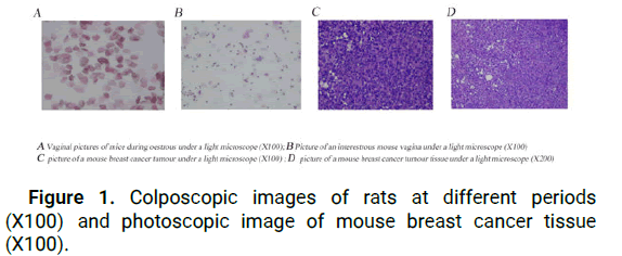
Figure 1: Colposcopic images of rats at different periods (X100) and photoscopic image of mouse breast cancer tissue (X100).
Establishment of a postoperative animal model of rat breast cancer: At 8 weeks after OVX, 4T1 cells in the logarithmic growth stage were taken and inoculated with 5 × 10⁵ cells in the right armpit of each mouse. Seven days later, the mice were observed and assessed. The epidermal tumours of mice were more than 0.3 cm in diameter and the mice were anaesthetized with chloral hydrate, followed by hair removal and routine disinfection around the tumours [11]. All tumour tissues were removed under aseptic conditions, with haemostasis and suture. Intraperitoneal injection of penicillin was given postoperatively. Mice with a good mental state, suture healing and no obvious tumour recurrence were selected one week after surgery, which was considered successful. Then, when 10% of the tumour tissue formalin was fixed for 48 hours, the tissue blocks were flushed with water for 6 hours and the tissue blocks were treated by an automatic dehydrator and embedded in paraffin. A 4 μm section was made and the tissue was glued onto the slide. On a baking sheet at 80°C for 4 h, HE staining was performed (Figure 2).
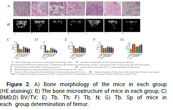
Figure 2: A) Bone morphology of the mice in each group (HE staining); B) The bone microstructure of mice in each group; C) BMD;D) BV/TV; E) Tb. Th; F) Tb. N; G) Tb. Sp of mice in each group determination of femur.
Experimental grouping and treatment
The experiment had 7 groups. Blank group: 10 BALB/c female mice at 8 weeks of age were given 20 ml/kg/d distilled water to 16 weeks of age and mice with body weights of 20 g to 25 g were randomly selected. Pseudocastration group: Adipose tissue similar to that of the ovary was removed from 8-week-old female mice and the other surgical procedures were the same as those of the model group. Eight weeks after surgery, 4T1 cells were inoculated and the tumour tissue was removed 1 week later [12]. One week after surgery, 10 BALB/c female mice weighing 20 g-25 g were selected. The remaining 5 groups: Eight weeks after the OVX operation, 4T1 cells were inoculated and tumour tissue was removed one week later. One week after the OVX operation, 50 BALB/c female mice weighing 20 g-25 g were selected and divided into 5 groups by a random method. The mice were divided into the model group (0.325 mg kg-1 d-1 letrozole), alendronate sodium group (1.3 mg kg-1 d-1 alendronate tablets) and the low-dose, medium-dose and high-dose (1.88, 3.76 mg kg-1 d-1 and 7.5 mg kg-1 d-1) Zuogui pill groups, with 10 animals in each group [13].
All groups were given the following treatments: the blank group and pseudocastration group, distilled water at 20 ml/kg/d; the model group (OVX+letrozole), letrozole at 0.325 mg kg-1 d-1; the alendronate sodium group, letrozole at 0.325 mg kg-1 d-1 +alendronate sodium tablets at 1.3 mg kg-1 d-1, the Zuogui pill high-dose group, letrozole at 0.325 mg kg-1 d-1+high-dose 7.52 mg kg-1 d-1, the Zuogui pill medium-dose group, letrozole at 0.325 mg kg-1 d-1+medium-dose 3.76 mg kg-1 d-1, the Zuogui pill low-dose group, letrozole at 0.325 mg kg-1 d-1+low dose 1.88 mg kg-1 d-1, the low-dose group had a clinical equivalent dose and the ratio of high, medium and low doses was 4:2:1. The drug was administered continuously for four weeks. The body mass of all mice was measured once a week, the dose was adjusted according to the body mass and the life and activity conditions were observed [14].
Specimen sampling and testing
All rats were anaesthetized with 10% chloral hydrate (0.03 ml/10 g body mass) and then fasted for one day before anaesthesia. Blood was extracted by eyeball extraction, centrifuged at low temperature (4°C, 3500 rpm/min, 10 min) and stored in the refrigerator at -80°C for use. After death, both lower limbs were removed and the surrounding soft tissues and muscles were removed. The right femur and tibia were placed in a sampling cup and fixed with 10% formalin. The bone density and trabecular microstructure in vitro were detected by Microcomputed Tomography (μCT) and the bone tissue morphology was observed by HE staining. The left femur and tibia were rinsed with normal saline, frozen in liquid nitrogen bottles and stored at -80°C. The expression levels of the Wnt3a, β-catenin and Runx2 proteins were detected by Western blots [15].
Serum oestradiol (E2) and the bone turnover markers BALP and P1NP were detected by ELISAs: According to the kit instructions, standard wells and sample wells were set. A control substance with different solubilities (50 μL) was added to each standard well. The sample diluent and the sample to be tested were successively added in the sample well on the enzyme label-coated plate. The sample was added to the bottom of the enzyme well, except for the blank well. The plate was sealed with sealing plate film and incubated at 37℃ for 60 min. The 20-fold concentrated washing liquid was diluted 20 times with distilled water for reserve use. The sealing plate film was removed, the liquid was discarded and the well was dried. Each well was filled with the washing liquid and then discarded after standing for 30 s. Colour developing agent A (50 μl) and then colour developing agent B (50 μl) were added to each well. The mixture was gently shaken and mixed and the colour developing process was carried out at 37℃ for 15 min. The final stop solution (50 μl) was added to each well to terminate the reaction (at this time, the blue turned to yellow), the blank well was adjusted to zero and the OD450 value of each well was detected within 15 minutes. The linear regression equation of the standard curve was calculated with the concentration and OD value of the standard substance. According to the correlation equation, the concentrations of serum oestradiol (E2), Bone Alkaline Phosphatase (BALP) and amino terminal propeptide of type I collagen (PINP) were calculated.
Histopathologic observation of femurs by HE staining: After the mice were killed, the attached tissue around the femur was cleaned and fixed in 4% paraformaldehyde for 10 days, decalcified in EDTA solution and then dehydrated, made transparent, waximpregnated, embedded and sectioned and then, bone tissue was stained by HE. The histopathologic morphology of femurs in each group was observed under a light microscope.
Microcomputed Tomography (μCT) bone microstructure test: Micro-CT bone microstructure measurements and analysis were performed in accordance with the guidelines of the American society for bone mineral salt research. The right femur fixed with 10% formalin was removed, local in vitro Bone Density (BMD) (g/ cm3) and trabecular microstructure of the right distal femur were detected by μCT scanning: Bone volume fraction (BV/TV, %), Bone Trabecular Thickness (Tb.Th, mm) and Bone Trabecular Number (Tb.N, 1/mm). When osteoporosis occurs, the bone volume fraction, bone trabecular thickness and bone trabecular number decrease, while Bone Trabecular Separation (Tb.Sp, mm) increases. The processed in vitro femur sample was placed into the scanning tube for scanning and the detection was carried out according to the standard operating specifications of the instrument. The scanning parameters were as follows: Voltage 60 kV, current 200 mA, exposure 400 ms and measurement accuracy 9 μm. Nrecon software was used to reconstruct the scan and the scanned data were imported into CTAn software. The growth plate was used as the reference level to draw the trabecular Region of Interest (ROI), and the thickness parameter was 0.5 mm. The femur region was manually selected to measure the trabecular structural parameters in the region of interest. The AC value of each part was calculated by the software and the BMD value (unit: g/cm3) was obtained by converting the AC value corresponding to the standard product scanned under the same conditions. Then, CTvox was used to construct a three-dimensional model to observe the changes in bone trabecular structure.
Determination of Wnt3a, β-catenin, Runx2 and protein expression in femur tissue by Western blot analysis: The femur tissue frozen at -80°C was removed, weighed, frozen again with liquid nitrogen and homogenized to extract protein. The total protein of femur tissue was extracted with RIPA lysis buffer according to the routine procedures of protein immunoblotting. After the protein was quantified by the BCA method, 20 μg was sampled, 10% SDS-PAGE was performed and the membrane was transferred by the semidry method and then sealed with 5% skim milk powder. Antibodies against Wnt3a (1:1000, Bioss), β-catenin (1:5000, Servicebio), Runx2 (1:8000, Servicebio) and β-actin (1:5000, Servicebio) were added and incubated overnight. After PBST washing, secondary antibody was added and incubated at room temperature for 2 h. After PBST washing, chemiluminescent reagent was added and the exposed film was developed and fixed with developing and fixing reagents. The optical density of the target strip was analysed by the alpha software processing system.
Statistical analysis
All data were presented with mean ± s.d and statistic analyzed by GraphPad Prism 9.5 software. Statistical diferences between groups were evaluated by one-way Analysis of Variance (ANOVA) followed by Least Signifcant Diference (LSD) test. The diferences were considered statistically signifcant when P value<0.05.
Results of vaginal smear tests
The results showed that the oestrous and interestrous periods in the blank group and pseudocastrated group lasted for 5 days. The other groups showed an interestrous period of 5 consecutive days, and more white blood cells and outer layer cells could be observed in the optical field, without regular changes in the oestrous cycle, indicating the success of the castration model and a good simulation of menopause.
HE staining of mouse breast cancer tumour tissue
HE staining showed diffuse solid growth, obvious atypia, large deep staining and pathological mitosis. The results showed that the postoperative model of breast cancer was successfully established.
Effect of Zuogui pill on bone morphology
Bone section observation under a light microscope showed that in the blank group, the bone trabecular structure was complete, the bone trabecular connection was uniform and the morphology of adipocytes and bone cells was clear. The basic structure of bone trabeculae was normal in the pseudocastrated group and the network structure was clear and orderly. In the model group, the bone trabeculae of mice were not continuous, the fracture was obvious, the number of fat cells increased and the diameter increased, indicating that the use of letrozole after castration aggravated the effect on bone. Compared with the model group, the high, middle and low-dose Zuogui pill groups and the alendronate sodium group showed no obvious poor bone trabecular structure and uniform bone trabecular connection, indicating improvements in the bone structure of each group. There was no significant difference between the high-, middle- and lowdose Zuogui pill groups and the alendronate sodium group, indicating that several groups of drugs improved osteoporotic bone changes.
Effects of Zuogui pills on Bone Mineral Density (BMD) in mice
The results were as follows: (1) Compared with that of the blank group, Bone Mineral Density (BMD) in the pseudocastration group and model group was significantly decreased and the difference was statistically significant (P<0.05). There was no significant difference between the model group and the pseudocastrated group (P>0.05), but the model group had a lower value than the pseudocastrated group, indicating that the use of letrozole tended to aggravate osteoporosis. (2) Compared with that of the model group, Bone Density (BMD) in the high-dose and medium-dose Zuogui pill and alendronate sodium groups was significantly increased (P<0.05). There was no significant difference in the low dose group (P>0.05). (3) Compared with the Zuogui pill groups, the alendronate sodium group showed no significant difference (P>0.05) and Bone Mineral Density (BMD) in the high-dose Zuogui pill group was better than that in the medium-dose and low-dose Zuogui pill groups (P<0.05).
Effects of Zuogui pill on the microstructure of the mouse femur
The results were as follows: (1) Compared with those of the normal group, Bone Volume/Tissue Volume (BV/TV, %), Trabecular Thickness (Tb. Th), bone volume fraction (Bone Volume/Tissue Volume, BV/TV,%), Bone Trabecular Thickness (Tb.th, mm) and Trabecular Number (Tb.N, 1/mm) were significantly decreased (P<0.05). However, Trabecular Spacing (Tb.Sp, mm) increased significantly (P<0.05). There was no significant difference between the model group and the pseudocastrated group (P>0.05), but the model group had a lower value than the pseudocastrated group, indicating that the use of letrozole tended to aggravate osteoporosis. (2) Compared with those of the model group, BV/TV, Tb.Th and Tb.N in the H-dose and M-dose ZGW and alendronate sodium groups were significantly increased, while Tb.Sp was significantly decreased (P<0.05). There was no significant difference in the low-dose Zuogui pill group (P>0.05). (3) There were no significant differences in BV/TV, Tb.Th, Tb.N and Tb.Sp of the alendronate sodium group compared with the H-dose ZGW group (P>0.05); the H-dose ZGW group had better values than the medium-dose and low-dose ZGW groups (P<0.05).
Effects of Zuogui pill on serum E2, BALP and PINP in mice
Effect of Zuogui pill on serum E2 in mice: The content of E2 in the blank group, pseudocastration group and model group was significantly different in pairs (P<0.01). There was no significant difference (P>0.05) in serum E2 content in all groups compared with the model group (high, medium and low Zuogui pill and alendronate sodium groups), but the content of serum E2 in the high, medium and low-dose Zuogui pill groups was higher than that in the alendronate sodium group.
Effects of Zuogui pill on the serum bone metabolic indexes BALP and PINP in rats: The contents of BALP and PINP were determined. The contents of BALP and PINP in the blank group, pseudocastration group and model group were significantly different (P<0.01). There were significant differences in serum BALP and PINP contents among the treatment groups compared to the model group (high, medium, low Zuogui pill and alendronate sodium groups) (P<0.01). The BAL and PINP contents in the high-dose Zuogui pill group were lower than those in the medium-dose and low-dose Zuogui pill groups (P<0.05) (Figure 3).
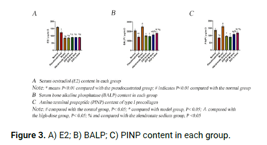
Figure 3: A) E2; B) BALP; C) PINP content in each group.
Effects of Zuogui pill on the expression of Wnt3a, β-catenin and Runx2 proteins in mouse bone tissue: The results were as follows: (1) Compared with those in the blank group, the protein levels of Wnt3a, Runx2 and β-catenin in the castration group and model group were significantly decreased (P<0.05). There was no significant difference between the model group and the castration group (P>0.05), but the model group had lower values than the castration group. (2) Compared with those of the model group, the protein contents of Wnt3a, Runx2 and β-catenin in the high-dose, middle-dose and low-dose Zuogui pill groups and alendronate sodium groups were significantly increased (P<0.05). (3) Compared with those of the high-dose Zuogui pill groups, the protein contents of Wnt3a, Runx2 and β-catenin in the alendronate sodium group were significantly different (P<0.05). The contents of Wnt3a, Runx2 and β-catenin in the high-dose Zuogui pill group were better than those in the medium and low-dose groups and the difference was statistically significant (P<0.05) (Figure 4).
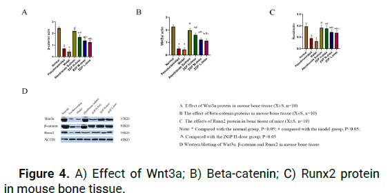
Figure 4: A) Effect of Wnt3a; B) Beta-catenin; C) Runx2 protein in mouse bone tissue.
Bone metabolism is a process with a complex dynamic balance that requires Osteoclasts (OC) to absorb old bone and Osteoblasts (OB) to form new bone. Only when the two are closely coordinated can human bone metabolism proceed normally. Oestrogen regulates bone metabolism in many ways, such as promoting apoptosis and reduction of OCs through direct and indirect effects and promoting proliferation and differentiation of OBs through cytokines. The ovarian function decline after menopause is caused by a sharp decline in oestrogen levels, as well as AI treatment of breast cancer caused by oestrogen synthesis disorders; the combination of these two factors results in low levels of oestrogen and the decrease of oestrogen level reduces OB activity, increases OC activity and destroys its dynamic balance. These changes promote bone loss and lead to Osteoporosis (OP).
OP belongs to the category of "bone impotence" in Chinese medicine. In "medical Jing Yi", it is stated that "kidney hides essence, essence generates pulp, and pulp generates bone", indicating that the kidney is closely related to bone. Kidney qi is sufficient and the bone is close. A lack of kidney qi leads to bone deficiency, namely, osteoporosis. Moreover, postmenopausal women suffer from Yin deficiency and blood deficiency, lack of viscera and bone loss, resulting in "bone impotence". Zuogui pill was originally from the "Jingyue Book" of the famous medical scientist Zhang Jingyue in the ming dynasty; it is a classic kidney tonifying prescription that reuses familiar land as the king, Jun tonifying kidney Yin and essence. This treatment uses C. officinalis to nourish liver and kidney and enhance the qi and has the effect of "invigorating pulp and Yang"; yam invigorates the spleen and Yin, nourishing the kidney and reinforcing essence; turtle shell and deer antler are used and the turtle parts nourish Yin and pulp, while the deer antler warms and strengthens kidney Yang, which are important components. Wolfberry has a tonifying effect on liver and kidney, invigorating blood, nourishing Yin but not Yin and promoting Yang and dodder seed "aids Yang solid drainage"; all these components are adjuvants. Together, these components tonify liver and kidney, strengthening the effect of muscles and bones. Zuogui Pill can enhance the level of hormone receptors by regulating hormone levels in the body or having hormone-like effects, thus affecting the growth and development of bones. Zuogui pill-containing serum could inhibit the expression of PTHrP and CXCR4 in tumour cells. In vitro, Zuogui pill-containing serum inhibited cytoskeletal F-actin polymerization. Studies have confirmed that Zuogui pill does not increase the level of oestrogen in the body but can protect against OP and other diseases caused by oestrogen deficiency. Studies have found that Zuogui pill and their combination could promote osteoblasts and inhibit adipocyte differentiation, effectively preventing the occurrence of PMOP.
Bone metabolic biochemical indexes such as the bone formation markers BALP (Bone-Specific Alkaline Phosphatase) and PINP (Procollagen I N-terminal Propeptide, type I procollagen amino terminal propeptide) can reflect the bone turnover rate in a timely and effective manner and can also be used to evaluate the efficacy of antiosteoporosis treatment. BALP levels have a linear relationship with OB and pre-OB activity and are a specific marker of bone formation for OB synthesis and secretion. High conversion metabolic bone diseases, such as Paget's disease, primary and secondary hyperparathyroidism and high conversion osteoporosis, all have increased BALP levels. P1 NP is produced by enzyme digestion and modification of type I procollagen, and its serum concentration reflects the synthesis level of type I collagen and can accurately reflect the formation of new bone. In addition, this molecule has high specificity and sensitivity in predicting the occurrence of OP, evaluating bone mass and monitoring anti-OP efficacy, and its detection is not affected by food, circadian rhythm, hormones and other interfering factors. Increased PINP is associated with accelerated bone turnover and bone loss. Therefore, PINP is an excellent novel clinical marker for bone metabolism-related diseases. The increase in BALP and PINP indirectly reflects the increase in bone loss and the change in clinical bone metabolic index occurs half a year earlier than bone density, so it is suitable for short-term observation indexes. Therefore, these two indexes can be used as reference indexes for the diagnosis and evaluation of curative effects after treatment.
The Wnt/β-catenin pathway is an important regulatory pathway of bone formation that plays a key role in bone homeostasis and bone repair. The mechanism of the Wnt/β-catenin pathway in bone metabolism has been continuously studied and has become a new hotspot in the pathogenesis and treatment of bone metabolic diseases. When extracellular Wnt binds with the membrane receptor Frizzled, dimers are formed through a series of interactions between membrane and cytoplasmic proteins, allowing β-catenin to accumulate in the cytoplasm. This molecule then enters the nucleus and cobinds to T-Cell-Specific Transcription Factor (TCF)/Lymphoid Enhancing Factor (LEF) to form a complex polymer that activates the downstream Runx2 gene. Osx gene expression is promoted to initiate target gene transcription, promote osteoblast differentiation and proliferation and exert its effects. This is the classic Wnt/β-catenin signalling pathway.
Wnt3a is the most representative signalling protein of the Wnt family and the main ligand of the Wnt/β-catenin pathway, which plays a role in regulating the function of osteoblasts. Inhibition of Wnt3a can lead to bone loss, decreased bone density and a decreased number of bone trabeculae. β-Catenin is an important downstream factor of the Wnt signalling pathway and plays an important role in cell signal transduction. When the Wnt signalling pathway is activated, β-catenin can bind to intracellular Transcription Factor (TCF/LEF) DNA to change the DNA structure, initiate downstream target gene transcription, and promote osteoblast differentiation and proliferation. In addition, experimental studies have found that the expression of the marker type I collagen, transcription factor osterix and osteocalcin in animal models with β-catenin gene deletion in the early stage of osteoblast differentiation is significantly reduced, which reduces the bone formation and mineralization of osteoblasts and leads to osteoporosis.
Runx2 is an OB-specific transcription factor found by Ducy P et al. through the study of OB-specific expression of the osteocalcin gene, which plays a key role in transcription. Runx2 is the main determinant of OB differentiation. This OB-encoding gene regulates the transcription of many genes. Runx2 plays an important role in osteoblast differentiation and bone formation and can influence osteoblast differentiation and bone formation through the classical Wnt/β-catenin signalling pathway, thus affecting normal bone development. Runx2 can enable bone marrow mesenchymal stem cells to differentiate and mature into chondrocytes and OBs, which has an important influence on bone repair and reconstruction. Many researchers have found that the Wnt/β-catenin signalling pathway can upregulate the expression of Runx2, a key bone factor and lead to OB differentiation. In OP fracture patients, the Wnt/β-catenin signalling pathway is involved in every stage of fracture healing, such as chondrocyte proliferation and differentiation, endochondral ossification and osteosynthesis. Callus resorption and remodelling are mediated by upregulation of Runx2 expression through the Wnt/ β-catenin signalling pathway. Runx2 deficiency causes bone loss in oestrogen deficiency. Oestrogen receptors in OBs bind Runx2, promote Runx2 transcription and increase the number of Runx2- expressing pre-OBs, thereby increasing bone mass and inhibiting the occurrence of OP.
The experimental results showed that the level of E2 in the pseudocastration group, blank group and control model group decreased successively (P<0.01), which indicated that the castration model was successful and the level of E2 in the control model group was the lowest, indicating that E2 was further reduced after the use of endocrine drugs. In this experiment, the serum oestrogen levels of mice in each group were measured and compared with those of the model group and there was no significant difference in serum E2 (P>0.05) among the treatment groups (high, medium, low Zuogui pill and alendronate sodium groups). Therefore, Zuogui pill in the treatment of secondary osteoporosis does not increase the risk of breast cancer. After treatment with endocrine drugs (letrozole), the bone tissue of mice with breast cancer after endocrine therapy was significantly damaged, and the serum contents of the bone metabolic indexes ALP and PINP were affected, indicating that the mouse postoperative osteoporosis model of postmenopausal breast cancer induced by endocrine therapy was successfully established. Zuogui pill intervention can improve bone morphology. Serum ALP and PINP levels of mice in the low, medium and high Zuogui pill groups decreased and were significantly different compared with those of the model group, which showed upregulated expression of Wnt3a, β-catenin and Runx2 proteins in bone tissue of mice with secondary osteoporosis after endocrine therapy. Activation and regulation of the protective mechanism of the Wnt/β-catenin pathway promote osteogenic differentiation, induce bone protection and protect against osteoporosis in mice with breast cancer after endocrine therapy (Figure 5).
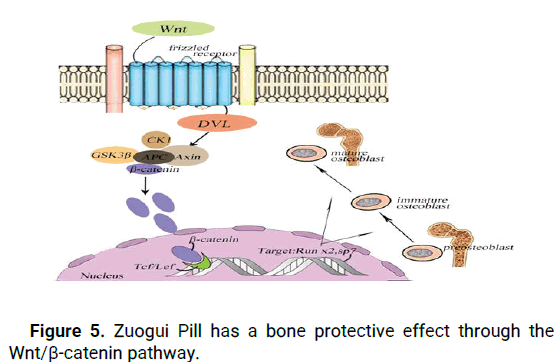
Figure 5: Zuogui Pill has a bone protective effect through the Wnt/β-catenin pathway.
In conclusion, this study found that Zuogui pill can affect bone formation, improve bone density, improve bone microstructure in mice, reduce serum BALP and PINP levels and upregulate the protein expression of Wnt3a, β-catenin and Runx2 in bone tissue of mice with secondary osteoporosis after endocrine therapy. Activation and regulation of the protective mechanism of the Wnt/β- catenin and Win/Runx2 pathways promote osteogenic differentiation and induce bone protection to prevent and treat secondary osteoporosis, especially in the treatment of secondary osteoporosis patients with breast cancer without increasing the risk of postmenopausal breast cancer induced by AI treatment. This study provides an experimental basis for treatment of bone metabolic diseases such as osteoporosis of breast cancer after clinical endocrine therapy. It is a safe, effective, low-cost and tolerable treatment method that is worthy of popularization and application.
We wish to thank American journal experts for scientific editing of this manuscript; we also thank the team of research of Inner Mongolia medical university.
Foundation and guidance: Liu Chunhui; Project design and manuscript writing: Ao Youguang; Index measurement and statistical analysis: Yao Lei, Zhuo La; Experimental validation: Jiang Zhaolei, Ma Jianchao, Shen Zhuorui.
This study was supported by the national natural science foundation of China (82060908); the health and family planning research program of Inner Mongolia autonomous region (201701051, 202202140); the 13th five-year plan project of Inner Mongolia department of education (NGJGH2018270); and Inner Mongolia medical university (NYGXTD20170).
The raw data for this article will be made available by the authors without undue reservation.
All animal experiments were approved by the committee of management and use of laboratory animals of inner mongolia medical university. (Ethics number: YKD202102110).
We declare that the publisher has the author’s permission to publish the relevant contribution.
The authors declare that there are no conficts of interest.
[Crossref] [Google Scholar] [PubMed]
[Crossref] [Google Scholar] [PubMed]
[Crossref] [Google Scholar] [PubMed]
[Crossref] [Google Scholar] [PubMed]
[Crossref] [Google Scholar] [PubMed]
[Crossref] [Google Scholar] [PubMed]
[Crossref] [Google Scholar] [PubMed]
[Crossref] [Google Scholar] [PubMed]
[Crossref] [Google Scholar] [PubMed]
[Crossref] [Google Scholar] [PubMed]
[Crossref] [Google Scholar] [PubMed]
[Crossref] [Google Scholar] [PubMed]
[Crossref] [Google Scholar] [PubMed]
[Crossref] [Google Scholar] [PubMed]
[Crossref] [Google Scholar] [PubMed]
Citation: Chunhui L, et al. "Study on Bone Protection Mechanism of Zuogui Pill on Osteoporosis Model of Breast Cancer Rats after Endocrine Therapy". J Biol Todays World, 2023,12(5), 1-6.
Received: 16-May-2023, Manuscript No. JBTW-23-98895; Editor assigned: 19-May-2023, Pre QC No. JBTW-23-98895 (PQ); Reviewed: 02-Jun-2023, QC No. JBTW-23-98895; Revised: 04-Aug-2023, Manuscript No. JBTW-23-98895 (R); Published: 01-Sep-2023, DOI: 10.35248/2322-3308-12.5.001
Copyright: © 2023 Chunhui L, et al. This is an open-access article distributed under the terms of the Creative Commons Attribution License, which permits unrestricted use, distribution and reproduction in any medium, provided the original author and source are credited.