Research - (2021) Volume 10, Issue 11
Background: Cleft Lip and Palate (CLP) are hereditary deformity of craniofacial structure. Temporomandibular disorder has number of clinical problems affecting muscles of mastication and its associated structures. There are several methods used to evaluate temporomandibular dysfunction (MRI, etc), Helkimo index is a settler in advancing indices, other indices used are CMI, MFIQ, FONSECA. The design of the following study was to evaluate the TMJ disorders in UCLP, if any, and compared with the non-cleft cases.
Methodology: Total 30 patients in age group of 16 to 20 years, will be selected from the patients coming to smile train Out Patient Department (OPD) of Orthodontics and Dentofacial Orthopaedics, Wardha. The total cases will be divided into two groups: a) Group I-unliteral cleft lip and palate cases b) Group II-non cleft class I cases Digital records of the patient (lateral cephalogram, photograph, models) will be taken and stored. Helkimo, MFIQ, CMI, Fonseca will be recorded on all patients participated in study. The Helkimo index will be taken to assess the presence and severity and MFIQ index will be taken to assess mandibular functioning in cleft cases based on the questionnaire.
Result: The dental apparatus (interdental relation of maxillary and mandibular dentition) plays an important role for the well-being of the temporomandibular joint. Trauma due to the occlusal instability has definite effect on the TMJ in long run. Cleft is always associated with inter jaw malocclusion and therefore TMDs in the cleft patient is thought to occur. As the treatment protocol for grown cases with severe skeletal malocclusion are orthognathic surgery. The TMJ correction remains untouched. It can be expected that altered TMJ anatomy can lead to TMDs in cleft lip and palate cases.
Conclusion: To convey the occurrence of TMDs in cleft due to the inter jaw malocclusion and its early treatment approach for TMJ deprogramming along with dental and surgical correction in CLP.
Cleft lip and palate • TMDs • Helkimo indices • MFIQ • CMI • Fonseca
Cleft lip and palate are the second most common congenital deformities of the craniofacial structure of infants which requires long-term functional and aesthetic rehabilitation [1]. After the primary surgery of lip and palate the healed fibrous tissue leads to restriction of the growth of the maxilla in all 3 planes (vertical, sagittal and transverse) which leads to maxillary post displacement as well as dysplasia [1]. The precedence of malocclusions in CLP patients is substantially high. Malocclusion basically occurrs in transverse plane; especially where the adjourn symmetry of the dental arches that can be clinically observed. They are also a prospective etiology of problems that are functional in nature, related to craniofacial structure. Hence, the patients with CLP are at risk of developing temporomandibular disorders due to mandibular disharmone [1].
Temporomandibular disorder embraces a number of clinical problems affecting the muscles of mastication, the Temporomandibular joint (TMJ) and its associated structures [1,2]. Mandible is secondarily affected part of the craniofacial apparatus in cleft. The relationship between the mandible and cranial base is important as it influences both sagittal, vertical facial disharmonies. The position of glenoid fossa is most likely to play an important role in the establishment of different craniofacial patterns. The fossa is affected by change in mandibular condyle position. Mandible which grows late based on the cephalocaudal growth gradients theory [3] is always under the confinement of the cranial base and maxilla. But due to three-dimensional constriction of maxilla, in the late growth spurt the mandible is gradually set free to grow forward downward depending on the altered growth and development of maxilla [3].
Contributing to the limited data on the effects of clefts on mandible. Few studies were performed in the department which evaluated and compared glenoid fossa morphology and depth in unilateral cleft lip and palate cases and the result suggested that, there was an increase in the depth and the width of the glenoid fossa and a decrease in the joint space, this may also influence the position of the articular disk [4-7]. In further studies, when the inclination of the condyle in the glenoid fossa was evaluated, it was found that, the altered inclination of the condyle results in a change in position of the articular disk which might further lead to the causation of TMD’s in UCLPcases [2,5,6,8]. The limitation of the said studies was, only the condyle and glenoid fossa was evaluated while disk position was not evaluated [8].
To evaluate of TMJ dysfunction various method are used like tomogram, magnetic resonance imaging, but Helkimo is a pioneer in developing indices. Which record severity by clinical evaluated [4]. Helkimo index measure the severity and pain of TMJ disorders and consist of three types: Anamesis, clinical, occlusal dysfunction [2]. This index is excellent means to allow check disease severity, measure effectiveness of TMD but the only limitation is in anamesis type anaylsis there mild and severe anaylsis but moderate option and for overcoming this limitation. Craniomandibular index is introduced to measure objective severity of mandibular movements, joint noise and muscle and joint tenderness using clearly defined criteria, simple clinical methods and ease in scoring [2,5].
As the dental apparatus and inter dental relation also plays an important role for the occurrence of TMD in cases without any craniofacial anomaly [9]. The inter-arch dental malocclusion is also assumed as a causative factor for factor TMD [3]. Meanwhile treating the CLP cases including orthopedic, orthodontic and orthognathic surgery [10,11]. The bite blocks used with the expansion devices act as TMJ deprogramming [10], can be expected to correct the TMD in the initial age, but this may not be true with all cases and these cases may show TMD in their later age to evaluate the occurrence of TMDs in the cleft [12-15]. The intention of doing this study was to evaluate the TMJ for disorders in UCLP, if any, and compared with the non-cleft cases.
Aim: To assess and comparison of temporomandibular joint for occurrence and severity of disorders in adult cases with unilateral cleft lip and palate and non-cleft class I.
Objective:
1. To evaluate the occurrence of TMD using Helkimo, MFIQ, CMI, Fonseca index in Non-cleft class I cases.
2. To evaluate the occurrence of TMD using Helkimo, MFIQ, CMI, Fonseca index in UCLP cases.
3. To compare severity of TMD between UCLP and non-cleft class I cases.
Hypothesis: Does the changed position of the mandible in craniofacial abnormalities like cleft deformities, the altered inclination of the condyle results in a change in position of the articular disk which might further lead to the causation of TMD’s in cleft lip and palate cases and does crossbite due hypoplastic maxilla predispose to TMD in unilateral cleft patients.
Source of the data
The subject to be studied will be selected from the smile train OPD and consent will be taken for the participation in the study, Department of Orthodontics, Sharad Pawar dental college, Sawangi.
Statistical analysis
Sample size formula for difference between 2 means
n=(2 α/22 × p × (1-P))/d2
2 α/2 is the level of significance at 5% i.e. 95% confidence interval=1.96
Confidence interval=1.96; P=prevalence of unilateral cleft lip and palate=0.7%=0.007
d=desired error of margin=5%=0.05
n=1.962 × 0.007 × (1-0.007)/0.0052=10.68=15 patients needed in each group
Study design
In this analytical study, a total 30 patients (UCLP and non-cleft class I), in age group of 16-20 years, will be selected. Approval form ethical community has been obtained (ref no. DMIMS(DU)/IEC/2020-21/9398).
The total cases will be divided into 2 groups:
a) Group I-unilateral cleft lip and palate cases
b) Group II-non cleft class I cases
Digital records of the patient (lateral cephalogram, photograph, models) will be taken and stored. Helkimo, MFIQ indices, CMI index, Fonseca index 13 will be recorded on all patients with cleft lip and palate. The Helkimo index will be taken to assess the presence and severity of TMD in cleft cases, while the MFIQ index will be taken to assess mandibular functioning in cleft cases based on the questionnaire and will be asked in their own language of understanding.
Inclusion criteria
1. UCLP cases of age group of 16-20 years
2. Class I bimaxillary, skeletal class I
3. All permanent teeth present
4. Based on cephalometric analysis
5. Class I
6. ANB-2°
7. Wit’s Appraisal -0-1 mm
8. Beta angle-27°-33°
9. Overjet -2-4 mm
10. Overbite-2-4 mm
Exclusion criteria
1. Patients who underwent any orthopaedic/orthodontic correction
2. Bilateral cleft lip/palate, only lip and anterior alveolus
3. Syndromic
4. Ankylosis cases
5. Cases with missing tooth, grossly carious teeth
6. Unilateral mastication, anterior cross bite, muscular dystrophy, bony deformities
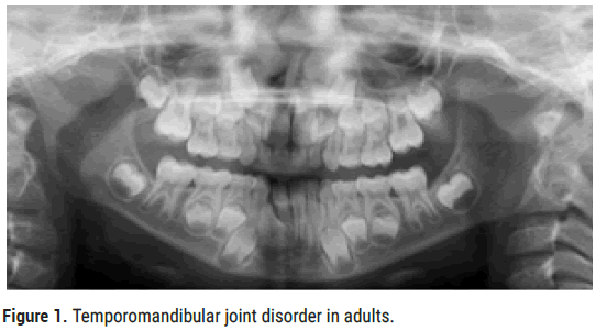
The dental apparatus (interdental relation of maxillary and mandibular dentition) plays an important role for the well-being of the temporomandibular joint. Trauma due to the occlusal instability has definite effect on the TMJ in long run. Cleft is always associated with inter jaw malocclusion and therefore TMDs in the cleft patient is thought to occur (Figure 1). As the treatment protocol for grown cases with severe skeletal malocclusion are orthognathic surgery.

Figure 1: 2021 Kumar N, et al. This is an open-access article distributed under the terms of the Creative Commons Attribution License, which permits unrestricted use, distribution, and reproduction in any medium, provided the original author and source are credited.
Ratnani et al. conducted study to estimate repair timing for palate when mandible is spatial oriented and morphology of mandible in relation to skull base and maxilla [4]. 20 surgically treated UCLP patients were categorized into two groups according to age. Study shows no significant effect on mandibular morphology and its position in UCLP cases with surgical repair timing of palate.
Patil et al. conducted a study which evaluated three dimensionally the condylar morphology, position and relation with glenoid fossa in unilateral cleft lip and palate cases treated with and without PNAM as compared to non-cleft cases, study consisted of 30 cases in the age group 9-12 year with 3 groups of PNAM, NON-PNAM, and NON-cleft cases [7]. Evaluation of the condylar morphology revealed that the height and the length of the neck of condyle was the longest in Class I non cleft cases and shortest in UCLP non PNAM cases. At the same time, the anteroposterior and mediolateral condylar widths were widest in UCLP non PNAM cases and narrowest in Class I non cleft cases. When the dimensions of the glenoid fossa were evaluated, it was found that its depth and the height of the eminence of the glenoid fossa were deepest and longest in UCLP non PNAM cases than in Class I non cleft cases. It was also observed that the width of the glenoid fossa was widest in Class I non cleft cases and narrowest in UCLP non PNAM cases. On evaluating, it was found that the anterior, posterior, superior and medial joint spaces were highest in Class I non cleft cases and lowest in UCLP non PNAM cases. And it also showed that lateral joint space was the highest in UCLP non PNAM cases and lowest in Class I non cleft.
khakhar et al. evaluated and compared the inclination of the condylar head with respect to glenoid fossa in cleft patients, class III, class I patients. Study consisted of 40 patients with 10 in each group of BCLP, UCLP, CLASS III, CLASS I [8]. Condylar head inclination angle was evaluated using 3D DVT method and results showed that in UCLP group the condylar inclination was more posteriorly than the non-cleft group.
Patil et al. studied the interrelationship between Skeletal and Dental Malocclusion in UCLP Cases based on Goslon Yardstick Scale [7]. Study consisted of 80 study model [Mixed=40, Permanent=40] using Goslon yardstick scale and their lateral cephalograms were evaluated for interjaw relationship. Significant interrater reliability was found for Golson yardstick scale. Goslon score-4 was found in 40% of study models of mixed dentition while Goslon score 2 and 3 each were found in 40% of study models in permanent dentition. Significant correlation was found between Goslon score and skeletal cephalometric parameters.
Sudheesh et al. studied the malocclusion status and treatment needs in patients with cleft and plate [9]. This was then compared and evaluated with non-cleft patients. This study consisted of 56 cleft lip and palate patients between 12-18 years of age and 168 non-cleft patients from the general population. The data was analysed with help of Dental Aesthics index (DAI). Around 51%-78% of cleft patients and 35.71% of non-cleft patients scored a DAI of 26-30 suggestive of definite malocclusion. This study thus concluded that most patients with cleft lip and palate exhibited severe malocclusion and thus will led TMDs [10-15].
To present the occurrence of TMDs in cleft due to the inter jaw malocclusion, early treatment approach of TMJ deprogramming along with dental and surgical correction in CLP can present the occurrence of TMDs in CLP. This study will help to evaluate TMD disorder using Helkimo, Cmi, Fonseca, MFIQ indices in CLP and compare it with non-cleft cases. The TMJ correction remains untouched. It can be expected that altered TMJ anatomy can lead to TMDs in cleft lip and palate cases.
Citation: Kumar N, et al. “Assessment and Comparison of Temporomandibular Joint for Occurrence and Severity of Disorders in Adult Cases with Unilateral Cleft Lip and Palate and Non-Cleft Class I: An Observational Study”. J Arthritis, 2021, 10(11), 001-003
Received: 08-Nov-2021 Published: 29-Nov-2021
Copyright: © 2021 Kumar N, et al. This is an open-access article distributed under the terms of the Creative Commons Attribution License, which permits unrestricted use, distribution, and reproduction in any medium, provided the original author and source are credited.