Case Report - (2017) Volume 3, Issue 2
Squamous cell carcinoma (SCC) conjunctiva is a rare cancer of the ocular surface with an incidence rate of 0.02 per 100,000 in high latitude areas to 3.5 per 100,000 at low latitudes near the Equator. This positive correlation between this SCC incidence and latitude indicates that exposure to ultraviolet radiation may play a role its etiology. The disease is more common in elderly and male patients, but may develop at a younger age, especially in association with xeroderma pigmentosum or immunodeficiency. Isolated squamous cell carcinoma of cornea is rare with few case reports. Here, we report two cases of conjunctival SCC, both in elderly males who presented with whitish painless mass encroaching the cornea. This mass was excised in both the patients. Histopathological examination confirmed the diagnosis of SCC conjunctiva.IOFF
Keywords: Squamous cell carcinoma; Conjunctiva
Squamous conjunctival carcinoma typically occurs on the bulbar conjunctiva, originating at the limbus, and often spreads onto the cornea. Even before the term ocular surface squamous neoplasia (OSSN) was introduced to encompass the spectrum of conjunctival and corneal intraepithelial neoplasia (CIN) and SCC [1-4], published series often included both intraepithelial and invasive squamous neoplasia. It is difficult to distinguish dysplasia, Carcinoma in situ (CIS), and SCC on clinical grounds alone. Histopathological examination is the confirmatory diagnostic modality. Treatment modalities include surgery, radiotherapy and topical cytotoxic agents. The prognosis is generally good if the conjunctival tumour can be completely removed. Chances of recurrence are high if the tumor grows into the eye and orbit. The mortality rate is generally are low about 4% to 8%.
A 70-year-old male presented to the OPD of Department of Ophthalmology with the complaint of brown colored growth in right eye for 2 months. This growth was progressively increasing in size and patient also complained of foreign body sensation (Figure 1). The growth was present in interpalpebral area in nasal bulbar conjunctiva extending 4 mm into limbus and cornea. On slit lamp biomicroscopy, a pinkish white, elevated, mass of 25 mm diameter was seen at nasal aspect of bulbar conjunctiva of right eye conjunctival congestion was noted adjacent to the lesion. Left eye was normal. There was no lymphadenopathy and systemic examination was within normal limits. All the routine investigations were within normal limits. The mass was excised and tissue was sent for histopathology.
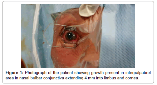
Figure 1: Photograph of the patient showing growth present in interpalpabrel area in nasal bulbar conjunctiva extending 4 mm into limbus and cornea.
A 60-year-old male presented to the OPD of the Department of Ophthalmology with a white mass in right eye associated with watering and diminution of vision since last 8 months. A 60-year-old male presented with history of progressively increasing swelling in the right eye for last six months associated with diminution of visual acuity in right eye. His general physical examination did not reveal any abnormality. On local examination, there was a growth measuring 15 mm × 12 mm located primarily in the temporal quadrant of the globe overlying the limbus and involving the temporal quadrant of the cornea (2 mm) (Figure 2). The growth was non-tender, fixed oval in shape and had gelatinous surface. Conjunctival congestion was seen and sentinel vessels were seen feeding the tumour. Visual acuity in his right eye was 6/18 and in left eye 6/9. Excision of the growth was carried out along with the healthy conjunctiva as freely as possible. The growth overlying cornea was peeled off easily. Superficial keratectomy was done. Postoperatively, the patient was put on 0.04% mitomycin C (MMC) eye drops four times a day along with antibiotic eye drops, non-steroidal anti-inflammatory eye drops and lubricating eye drops four times a day. MMC 0.04% eye drops were applied four times a day for 14 consecutive days followed by 7 consecutive days off MMC. The treatment cycle (14 days on MMC and 7 days off) was repeated for three cycles. Adjacent to the lesion conjunctival congestion was present. Left eye was normal. Tissue was sent to department of histopathology.

Figure 2: Photograph of the patient showing a growth measuring 15 mm × 12 mm located primarily in the temporal quadrant of the globe overlying the limbus and involving the temporal quadrant of the cornea (2 mm).
Both the biopsies showed similar histopathological features. Sections studied show nests of squamous epithelial cells arising from the squamous epithelium and extending into the sub epithelial tissue. The malignant cells were large with abundant eosinophilic cytoplasm and a large vesicular nucleus (Figures 3a and 3b). Keratin pearls were present. A diagnosis of moderately differentiated squamous cell carcinoma was made in both the cases.

Figure 3a and 3b: Microsections examined show nests of squamous epithelial cells arising from the squamous epithelium and extending into the subepithelial tissue.
Squamous cell carcinoma (SCC) arising from conjunctiva is an uncommon disease world wise, the incidence of which varies from 0.02 to 3.5 per 100 000 [1,2]. Since the 1980s, the number of patients presenting with squamous cell carcinoma of the conjunctiva has been increasing exponentially [2,3]. This lesion has a multi factorial etiology with interplay of several factors like exposure to ultraviolet radiation, various chemical carcinogens and viral infections (HPV), however role of individual agents is not well understood. OSSN predominantly occurs in elderly males with an average age of 56 years [1]. This may be because jobs that tend to expose people to sunlight over long periods of time are commonly done by men. People with AIDS have an increased risk of developing certain cancers, including SCC of the conjunctiva
This tumour often appears in the area closest to the nose or temple. The growth may present as a white, flesh-coloured or red patch or a rounded, elevated growth or growths that have a gel-like appearance. It can cause eye irritation of the effected eye or chronic conjunctivitis (inflammation of the conjunctiva). SCC should be considered in cases of conjunctivitis that last longer than 3 months. These tumours may mimic benign conjunctival degenerations and present as slowly growing lesions. SCC may coexist with pinguecula and pterygia [5]. Diffuse tumours in older patients may be misdiagnosed as a chronic unilateral conjunctivitis [6,7]. The main barrier to prevent invasion is Bowman’s layer [8]. Rarely the initial lesion appears on palpebral conjunctiva. The lesions neglected for a long time may spread into the globe or orbit.
The importance of HPV types 16 and 18 in the pathophysiology of conjunctival carcinoma is uncertain. There are some studies that indicate the association of Human Papilloma Virus-16 (HPV-16) with some cases of bilateral conjunctival dysplasia. Around 77% of cases have had immune histogenic evidence of these viruses [9]. Also, cutaneous and visceral malignancies in association with SCC have been reported. Erie and associates reported 13% of patients had a history of malignant skin tumours [10].
The treatment options for conjunctival epithelial malignancies include excision of the tumour removal with or without cryotherapy, radiotherapy and topical chemotherapy [10]. Recurrent conjunctival squamous cell carcinoma means that the cancer has come back after it has been treated. It may recur in the same location as the original cancer or it may recur in another part of the body (metastatic conjunctival squamous cell carcinoma). The status of surgical margins. The reported incidence of recurrence after initial treatment is quite variable. Previous reports reveal high recurrence rates after simple surgical excision around 24% to 50%. Invasive disease may cause intraocular and orbital involvement then exenteration may be needed. Reconstruction of the conjunctiva may be required, depending on the amount of tissue removed. This may include using grafts of nearby tissue for reconstruction.
Conjunctival squamous cell neoplasms can cause significant morbidity. Early diagnosis and intervention can prevent major eye damage. Surgery with excision of margins and adjuvant cryotherapy has a very high success rate. Technical modification may sometimes be necessary to reduce damage. A long follow up is necessary to prevent the morbidity.