Case Report - (2023) Volume 8, Issue 5
Background: Plantar keratodermas are a diverse group of hereditary and acquired keratodermas characterised by hyperkeratosis of the skin of the soles. Hyperkeratotic eczema of hand/feet typically chronic and refractory to therapy is still a poorly understood entity associated with pain mobility impairment and functional limitations.
Case report: This case report is on an idiopathic plantar hyperkeratosis in a 55 year-old Chinese female with no prior dermatology history. The patient presented to the skin clinic with a recurring left plantar dermatitis with frequent bleeding and skin breakdown 5-years ago. It then spread to both plantar especially on the lateral border of both feet about 3 years ago. Initial laboratory examination for fungal and patch tests returned with negative findings. On the 4th year a punch knife biopsy for malignancy was performed and results were negatives. Hyperkeratosis followed by skin breakdown continues until today.
Conclusion: Patient self-management approach to comprehensive care is needed for plantar-hyperkeratotic eczema to complement medical management as PKK takes a chronic form of illness. The medication regimes starting with antifungal antibiotic-steroid cream keratolytic agent and a course of acitretin tablets with regular outpatient wound-care dressing is insufficient for this case. Relief was unsustainable and symptoms recurrences impacted quality of life. Collaborative patient self management to properly engage patient in partnership with self-care strategies to target at resolution with minimal adverse effects (in terms of scarring crusting skin breakdown and bleeding) are needed with additional consideration of other personal-environmental causal factors.
Dermatitis • Keratodermas• Wound-care • Patient self management
Palmoplantar Keratodermas (PPKs) is a cluster of skin disorders characterized by abnormal thickening (hyperkeratosis) of the skin on the palms and soles and often involves greater than 50% of the involved surfaces areas. Broadly classified as either hereditary or acquired PPK have numerous underlying causes which includes drug related malnutrition associated, chemically-induced systemic disease related keratoderma dermatoses related infectious malignancy associated and idiopathic BMI and obesity-related. As a somewhat neglected entity these hyperkeratotic eczemas are characterized by chronic scaly hyperkeratotic erythematous plaques on the plantar and/or palmar surfaces. Treatment option for PPK include topical keratolytic-salicylic acid repeated scalpel debridement topical retinoids topical psoralen plus UVA and/or topical corticosteroids, with wound care dressings in serious skin breakdowns. The presence of pain itch and skin break downs and frequent clinic visits can lead to considerable psychological distress. Hyperkeratotic eczema of hand/feet typically chronic and refractory to therapy is not a poorly understood entity but it’s often difficult to manage with unclear etiologies and when prolonged can negatively reduce patients’ quality of life-since they are associated with pain and functional limitations. This case on a Palmoplantar Keratoderma (PPK) highlighted that traditional management of keratosis with a specialist instructing a passive patient to follow instruction in a chronic condition is insufficient and calls for engaging an active involvement of patient in symptom managements and chronic disease management to approach the condition holistically. Informed consent was obtained from the patient for publication of this case report and accompanying images [1].
This case report describes a case of simple eczema developing into a chronic plantar keratosis of at least five years period affecting both sole and impacting walking ability. The purpose is to report the clinical decision-making but also to highlight the need partner with patient for collaborative patient selfmanagement care of a chronic plantar hyperkeratotic eczema patient especially in a traditional medical model hospital setting. Quality of life with chronic Palmoplantar Keratodermas (PPKs) are dependent not only on effective management of the medical aspects (on the location area and extend of spread and symptom control where underlying cause is idiopathic) but also on non-medical aspect of roles and relationship and emotional management as chronic conditions affect participation of daily living performance. The report is guided by the care checklist of information to include for case report writing [2].
A 55-year-old woman presented to a skin clinic with a 4 weeks history of localised rash on her left plantar (distal sole). She was diagnosed with contact dermatitis (actopic aczema) in June 2014. Patient is 65 kg 5 feet 2 inches tall with BMI of 26 and past history of hyperlipidemia. The patient described the rashes as dry weeping painful and burning with weekly skin breakdown and lacerated wounds. She does not have any recent illness fever and unsure of any environmental exposure. Home treatment included epsom salt bath bodyshop foot spa treatment and moisturisers of several types including squalene oil. The eczema worsened and spread to both plantar with complaints of cycles of skin dryness scaling and weeping rashes around the middle posterior soles and along both lateral borders of the feet with at least one skin breakdown with/without bleedings every 1-2 weeks initially [3].
Table 1 presents her skin specialists’ consultation notes and clinical decision makings. A fungal test of scrapped skin was performed at six months after the onset but it did not show any fungal infection. She was later prescribed a patch test but findings were negatives. Clinical examination showed well demarcated erythematous scaly and hyperkeratotic plaques with scattered pustular lesions on bilateral planate over the posterior sole and along the lateral borders. She was subsequently diagnosed as, hyperkeratotic xerosis. Medications prescribed for her included steroid cream antibiotic and moisturiser with followed up between 2-6 months intervals [4].
| Skin specialists’ clinical decision making | Patient self management | |
|---|---|---|
|
||
| 30/6 | Underlying dyslipidaemia with no known allergies or family history of atopy peeling skin over left heels × 2/12, not itchy seen a GP in March 2014 and given injection (antibiotics), betamethasone cream, Johnson baby oil. No change in footwear, using cream from body shop × 1 week. Xerotic, thickened skin, with skin peeling over heels ++ Imp-Heel Eczema-Rx urea cream | - |
| 28/11 | Cracked left heel, scaling, desquamation × 3/12 skin scraping sent (-negative result). Rx: 10% urea cream. Left heel-scaling, desquamation × 3/12 | |
| 19/12 | Scaling and erythematous patches over left sole no fungal element seen. Rx: Aqueous cream and miconazole 2% cream | |
2015 |
||
| 17.11 | No palmar involvement no oesophageal motility problems, no weight loss/night sweats/fever. (Dx: Left Plantar hyperkeratosis), Rx: Emulsifying ointment mupirocin 2% ointment clobetasol propionate 0.05% ointment at night to red areas | Change in footwear-soft shoes and clean sock daily at work |
2016 |
||
| 3/6 | Left and right plantar presented with well demarcated erythema involving instep, with complaints of itch over bilateral plantars, on-off over past 2 years. (Imp: Tinea pedis and plantar eczema), Rx: Aqueous cream, hydrocortisone 1% cream, cetirizine 10 mg OD, itraconazole 200mg OD × 2/52, ketoconazole 2% cream, KMnO4 1:10000 soaks | KMn04 soak daily, daily cling foil wrapped after moisturising both feet |
| 6/9 | Bilateral scaly feet with moccasin-like pattern and with hyperkeratosis previous scraping was tested negative for fungal infection. Patch test done-no significant results. (Dx: Hyperkeratosis of feet >6/12 ± secondary fungal infection), Rx: Repeat itraconazole 200 mg OD × 2/52, miconazole 2% cream LA BD, review in 2 weeks KIV acitretin | - |
| 1/11 | Improved subjectively with softened skin, (Dx: Plantar hyperkeratosis) Rx: Acitretin 25 mg 3 × week reduced (to reduce S/E dryness of lips) | |
| 13/12 | Desquamation on feet-better than before Rx: Acitretin 25 mg 3 x week, vaseline to lips, emulsifying ointment to soles | |
2017 |
||
| 7/2 | Acitretin 25 mg 2 × week to reduce peeling of plantar skin salicylic acid 10% at night | Use of slipper at home. Avoid irritants |
| 7/3 | Emulsifying ointment, acitretin 25 mg 2 × week, salicylic acid 10% at night | Moisturises both feet at night and cling foiled till morning |
| 23/11 | Stopped acitretin a year ago, after some improvement, then recurred, desquamation ++, some pain 2/7 ago, plantar hyperkeratosis with cracks, Rx: Betamethasone dipropionate 0.05% salicylic acid 3% (beprosalic) ointment twice daily, emulsifying ointment twice daily, fobancort/fucicort cream twice daily to cracked areas. Dx: Plantar hyperkeratosis? eczema | - |
2019 |
||
| 17.5 | Plaques with cracks, left hip wart, Rx: Aqueous cream, emulsifying oil, beprosalic, cryo LN2 2 x 10s; Dx: Plantar hyperkeratosis, wart | Soft padded shoe, prevent skin breakdown |
| 4/7 | Punch biopsy performed. (result-negative) | - |
| 6/8 | Reducing scaly plaques on soles; Rx: Emulsifying, beprosalic, betamethasone dipropionate 0.05% ointment, fucicort cream LA BD to cracks. Dx: Eczema TRO allergic contact dermatitis | Home remedy of garlic, (with squalene oil from GP) |
Table 1. Clinical decision making and patient self management.
On the second year after initial diagnosis, the hyperkeratosis spread to the right feet, and she was diagnosed as bilateral plantar hyperkeratosis. On examination, both plantar present with chronic scaly erythematous plaques. She was started on keratolytic therapy, topical corticosteroid and moisturizers. In 2017, the plagues were crusting and with occasional scattered pustules and bleeding episodes. She was prescribed a course of oral retinoid therapy. In 2018, skin breakdown was less frequent at once every 2-3 months if self-care of the feet was consistent. In 2020, bleeding and skin breakdown was once in 3-6 months-patients claimed a regimented moisturising and nightly cling-foil both feet helps control the skin dryness. Her medications includes steroid, antibiotic cream and moisturiser [5].
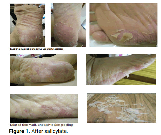
This case of idiopathic eczema, initially presented to the skin clinic with only a small red spot (2 inches by 1 inch) on the left posterior plantar. Patient received a diagnosis of contact dermatitis on June 2014 and the prescribed steroid (betamethasone cream) did not resolve the condition. A fungal test patch test and punch biopsy all returned with negative findings. The exact cause of plantar hyperkeratosis is unknown but genetic and environmental factors obesity-related inflammation and inflammatory cytokines interleukin may play a role in the pathogenesis of these skin disorders (Figure 1).

Figure 1: After salicylate.
Investigation
The differential diagnosis includes contact dermatitis fungal allergy and bacterial infection. On initial examination contact dermatitis was considered because the rash was localised and well demarcated but the rashes did not resolve with prescription of antihistamines, emollients and topical steroids. On subsequent visits a fungal infection which are typically asymmetric was considered (when presentation was only on the left foot)-but the antifungal drugs did not resolve the symptoms [6].
Follow-up and management
The PPK persisted with period of exacerbation and remissions-with episodes of erythroderma hyperkeratosis and skin peeling and pustules which coalesce to resolve in the next few days-appearing as brown macules and fissures with burning pain affecting activities of daily living (especially walking). On several occasions the open skin lesions have resulted in several secondary bacterial infections with patients reporting visits to GPs with symptoms of fever purulence for a bacterial complication rather than bacterial origins. Patients were given MC to rest and a course of antibiotic with fucidin cream and moisturiser from the GPs [7].
The treatment options for PPK vary from topical to systemic therapy with topical corticosteroids and wound care dressings oral retinoids (25 mg daily acitrecin ovr 4-8 weeks) keratolytics (salicylate acids) as firstline therapy. Systemic retinoid therapy has been recommended for chronic hyperkeratotic palmoplantar dermatitis with oral acitretin for treatment of chronic hyperkeratotic eczema and with one study reporting results superior to topical corticosteroid and keratolytic therapy [8].
Conservative daily intervention to maintain remission include avoidant of dryness avoidant of irritants (with daily wear of slipper at home and soft shoes at work) and skin emollients. Patient also started to step-up self-care with a regimented regime of i) nightly moisturizer (cling foil wrapped), ii) fucidin cream (if weeping rashes ocurrs), iii) trimming off excessive hardened skin peels and iv) home remedy of garlic paste with squalene oil over both feets 1-2 times daytime in a week or when the feet is especially dry [9].
Outcome
The cling foil method of both feet, kept the feet well moisturised even in the air-conditioned bedroom. Patient had less episodes of skin breakdowns (from twice a month breakdown to once in a month or in two months). Hyperkeratosis have been treated effectively with keratolytic therapy but acitrecin was proven superior to topical corticosteroid and keratolytic therapy. Both treatments showed improvement in terms of less flare up of symptoms (skin breakdowns, bleeding and pain and affected mobility) especially in the initial one month, but it did not improve the quality of life and the acitrecin had some adverse effects not tolerated well by the patient, who experienced dry mouth/lips/tongue affecting her communication. The patient was a lecturer and was very conscious of the mouth dryness. She was assured that the use of retinoids over the long term appears to be safe but was still anxious and dosage was lowered from (10 mg) daily to 3 x a week. Clinical and laboratory monitoring (serum lipid and hepatic profiles) were ordered to detect any significant elevation in lipids or liver enzymes but no abnormalities was observed. After 5 months patients asked to stop the acitrecin and was placed back on the usual steroidal cream (with fucidine cream if skin break down). A prescription of betamethasone dipropionate 0.05% salicylic acid 3% (beprosalic) ointment twice daily emulsifying ointment twice daily fobancort/fucicort cream twice daily to cracked areas was the latest regime. Apart from scalpel debridement systemic retinoids and use of salicylic acid a newer non conservative modality using split-thickness skin graft have successfully treated plantar (corn) hyperkeratosis with shorter recovery time. However non-medical care aspects have also shown results such as proper shoe wear and obesity related inflamatory skin disease must be intervene with weight management self care. Obesity is one of the important causal factors of many inflammatory skin diseases but environmental factors are also significant triggers requiring complex behavioural changes and interventions not just drug prescriptions [10].
Patient self-management
With chronic and systemic condition patient selfmanagement with good evidence of effectiveness should be pursued in management of complex PKK cases. For clinicaleconomic reasons the number of persons living with chronic PPK conditions may represent a new significant public health issue and a registry may be timely to provide data for research into this area to inform intervention. Linking in patient-responsibility with selfmanagement approach could present a promising strategy for chronic skin conditions. With healthcare moving beyond traditional patient-education to engage individuals to solve problems related to their chronic skin illnesses through various self-management processes to build up patient self-efficacy and empowerment and resulting in health-behaviour improvement better functional health status quality of life and also psychological well-being. In fact recent studies also show that obesity is a major risk factor for the development of inflammatory skin diseases like eczema and atopic dermatitis. Higher BMI percentiles have been found associated with higher odds of eczema compared with lower BMI suggesting weight management in patients with ezcema. One study even suggest that older people with hyperkeratosis are linked with poor footwear and low frequency of foot health checks [11-13].
Therefore, more efficacious treatment is needed to ensure better comprehensive management and effective symptom management to reduce the associated pain mobility impairment and interference with function and quality of life. In chronic cases of more than 6 months of antibiotics usage merely controls (and do not cure) the condition and treatment can be life-long. With such long duration patient involvement must be optimized with chronic disease self-management, a matured science with good evidence of improvement in quality of life to target long-term adherence for therapeutic regimens (prescribed medication changes in dietary and exercise practices for weight and overall health too) that can improve functional status and health outcomes [14].
Patient perspectives
The use of keratolytic drugs was painful as it causes dilated thin walls, requires and skin peelings was excessive and self care debridement was time consuming to prevent open wound. A ritual of cling-foiled wrapping of feet with moisturiser appears to be the best self-management strategy in controlling the hyperkeratosis [15].
Conservative management of the patient’s hyperkeratosis eczema (typically anti-fungal steroid antibiotic with a course of acitretin tablets and outpatient wound-care to debride the hyperkeratosis) did result in relief but it was intermittent and unsustainable. Conservative management of the hyperkeratosis must be stepped up with early patient self-management to ensure partnership in a comprehensive care aiming at minimal adverse effects and complete resolution (of diffuse scarring crusting skin breakdown recurrences and bleeding) longer interval for chronic recurrences but also to consider potential personalenvironmental triggers. Hyperkeratotic eczema of feet typically chronic and refractory to therapy is still a poorly managed entity and associated with chronic pain prolonged mobility impairment and functional limitations which warrants intervention from a chronic disease management.
Written informed consent was obtained from the patient for publication of this case report and accompanying images.
The author declares no potential conflict of interest with respect to the research, authorship and/or publication of this article.
[Crossref] [Google Scholar] [PubMed]
[Crossref] [Google Scholar] [PubMed]
[Crossref] [Google Scholar] [PubMed]
[Crossref] [Google Scholar] [PubMed]
[Crossref] [Google Scholar] [PubMed]
[Crossref] [Google Scholar] [PubMed]
[Crossref] [Google Scholar] [PubMed]
[Crossref] [Google Scholar] [PubMed]
[Crossref] [Google Scholar] [PubMed]
[Crossref] [Google Scholar] [PubMed]
[Crossref] [Google Scholar] [PubMed]
[Crossref] [Google Scholar] [PubMed]
[Crossref] [Google Scholar] [PubMed]
Citation: Loh SY. "Patient Self Management Rehabilitation for Plantar-Hyperkeratotic Eczema: A Case Report". Med Rep Case Stud, 2023, 8(5), 1-4.
Received: 24-Apr-2020, Manuscript No. MRCS-23-9724; Editor assigned: 29-Apr-2020, Pre QC No. MRCS-23-9724 (PQ); Reviewed: 13-May-2020, QC No. MRCS-23-9724; Revised: 18-Jul-2023, Manuscript No. MRCS-23-9724 (R); Published: 15-Aug-2023, DOI: 10.4172/2572-5130.23.8(05).1000252
Copyright: © 2023 Loh SY. This is an open-access article distributed under the terms of the Creative Commons Attribution License, which permits unrestricted use, distribution and reproduction in any medium, provided the original author and source are credited.