Case Report - (2017) Volume 3, Issue 3
Sarcomas of the omentum are rare. In this article, we report the case of a sarcoma of the omentum which was diagnosed and treated at the Joliot Curie Cancer Institute of Dakar. The patient was a 52-year-old woman with an abdominal mass that was mistaken for an ovarian tumor on clinical examination and on imaging. Surgical exploration helped find a sarcoma of the great omentum and treat the disease. Adjuvant treatments depend on the prognostic factors that need to be defined.
Keywords: Sarcoma; Omentum; Surgery; Prognosis
The diseases of the peritoneum are numerous. They are dominated by nonspecific infections and tuberculosis. Benign tumors are rare. Secondary malignancies are common and are dominated by peritoneal carcinomatosis of gynecological and digestive origins [1]. Soft tissue sarcomas are primary malignant tumors of mesenchymal or mixed natures that develop at the expense of soft tissue [2]. Clinically and in medical imaging, it turns into tumors that are often voluminous. This is a heterogenous histologic tumor with more than 100 subtypes [3]. It usually sits on the lower and upper limbs, retroperitoneum, mediastinum. Exceptionally, the epiploon, an essentially fatty tissue, is the location of sarcomatous diseases. We report a case of a primary omentum sarcoma treated at the Joliot Curie Institute in Dakar.
The patient was a 52-year-old female with no specific medical history. She noticed a painless abdominopelvic mass that rapidly increased in volume in a 3-month period, associated with vomiting and other symptoms. Clinical examination found an enlarged, distended abdomen. The mass was located in the umbilical area. It had no peripheral lymphadenopathy. A pelvic touch found a right laterus mass. The level of CA 125 was 29.29 IU/ml. The abdomino-pelvic computed tomography showed a mass of 160 mm × 90 mm occupying the abdominopelvic cavity with tissular density, hyper-vascularity, and lobulated contours (Figure 1). Surgery found a voluminous tumor which was about 20 cm long, of cerebroid aspect and soft consistency, that developed exclusively at the expense of the great omentum. The intra and retro peritoneal organs and the internal genital organs were normal. Abdominal adenopathies were not found. An intragastrical omentectomy was carried out (removed) with the tumor (Figures 2). Pathology of the surgical specimen showed a proliferation of spindle-shaped malignant tumors or atypical ovoid cells, sometimes presenting mitoses, and clustered in bundles separated by vascular spaces in favor of a spindle cell sarcoma (Figures 3). Cisplatin and Adriamycin chemotherapy was initiated for 6 cycles. Tolerance was good. After 30 months of follow-up, the patient was clinically and radiologically asymptomatic.
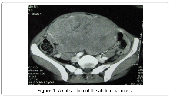
Figure 1: Axial section of the abdominal mass.
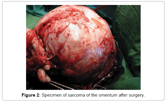
Figure 2: Specimen of sarcoma of the omentum after surgery.
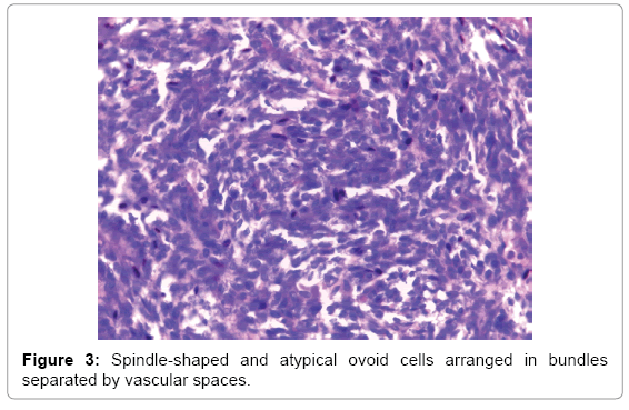
Figure 3: Spindle-shaped and atypical ovoid cells arranged in bundles separated by vascular spaces.
Secondary tumors of the omentum are common in peritoneal carcinomas; they usually are of digestive or gynecological origin [4]. Soft tissue sarcomas account for 0.5% to 1% of cancers in adults [2]. Sarcomas of the omentum are rarely described, and a limited number of cases have been reported [5,6].
The age of occurrence of sarcomas is variable. In children there is a high incidence of embryonic sarcomas from various sites. A second peak occurs in late adolescence. In adults, the most frequent localizations are extra-abdominal. Intra-abdominal sarcomas are retro peritoneal and sometimes mesenteric. They can also have developed at the expense of the stomach, intestine or pancreas in the context of Gastrointestinal Stromal Tumors (GIST). Localization in the omentum makes it difficult to identify a predilection for age and sex [4,7].
Symptomatology is dominated by abdominal pain and distension. In women, the first diagnostic is always that of an ovarian tumor before GIST, mesenteric tumor and tumor of the retroperitoneum [8,9].
The circumstances of the discovery of sarcomas are most often clinical. There is no screening program for abdominal and ovarian tumors in women. Patients are therefore seen at a late stage. Size is an important prognostic argument [10]. Computed Tomography is the first-line of examination, while Magnetic Resonance Imaging is the reference. Positron Emission Tomography-Computed Tomography (PET-CT) can be used to assess residuals lesions after surgery and early metastasis [11].
Surgical exploration is the rule when dealing with a well-defined abdominal tumor without peritoneal carcinosis, ascitis and metastasis [12]. Surgery should take all the structures infiltrated by the tumor with wide margins [13,14]. Margins in soft tissue sarcomas are variable. The desired margins are limited by the proximity of noble organs such as the pancreas, retroperitoneal and mesenteric vessels [15]. The presence of adenopathies in soft tissue sarcomas is an important prognostic event [16].
Pathology often finds a voluminous mass that is very limited, irregular, and cerebroid as observed in our case. We also noticed solid white parts, and areas of necrosis. Microscopy and immunohistochemistry pose the diagnosis of sarcoma and guide the molecular profile [6]. The most common histological type in the omentum is liposarcoma [3,5].
Adjuvant treatments improve local control. The use of radiotherapy is very limited for intra-abdominal tumors. The most used adjuvant treatment is chemotherapy [7,17]. Current hopes for adjuvant medical treatments are based on the use of targeted therapies taking into consideration the tumor molecular signature [18].
Primary sarcoma of the great omentum is rare. Surgical exploration is of great importance to the diagnosis, in addition to clinical and imaging explorations. The histological forms are varied. Liposarcoma is the most commonly described type. The discovery of new cases will make it possible to establish more prognosis and better adjuvant therapeutic choices.
Primary We thank the pathology laboratory for providing us with the pictures of the pathological examination.